Tropomyosin

|
WikiDoc Resources for Tropomyosin |
|
Articles |
|---|
|
Most recent articles on Tropomyosin Most cited articles on Tropomyosin |
|
Media |
|
Powerpoint slides on Tropomyosin |
|
Evidence Based Medicine |
|
Clinical Trials |
|
Ongoing Trials on Tropomyosin at Clinical Trials.gov Clinical Trials on Tropomyosin at Google
|
|
Guidelines / Policies / Govt |
|
US National Guidelines Clearinghouse on Tropomyosin
|
|
Books |
|
News |
|
Commentary |
|
Definitions |
|
Patient Resources / Community |
|
Patient resources on Tropomyosin Discussion groups on Tropomyosin Patient Handouts on Tropomyosin Directions to Hospitals Treating Tropomyosin Risk calculators and risk factors for Tropomyosin
|
|
Healthcare Provider Resources |
|
Causes & Risk Factors for Tropomyosin |
|
Continuing Medical Education (CME) |
|
International |
|
|
|
Business |
|
Experimental / Informatics |
Overview
Tropomyosin, along with troponin, regulates the shortening of the muscle protein filaments actin and myosin. In resting muscle fibres, tropomyosin is displaced from its normal binding groove by troponin. This displaced conformation of tropomyosin prevents the binding of myosin heads, thereby inhibiting muscle contraction. Under normal stimulation to muscle fibers, Ca2+ ions are released from the sarcoplasmic reticulum of the myocytes which causes troponin to release its hold on actin, allowing tropomyosin to return to a normal conformation which allows myosin heads to walk along actin filaments and thereby facilitating muscle contraction.
Functional Characteristics
Sliding filament theory
Tropomyosin is an alpha helical coiled coil protein dimer that binds end to end along F actin filaments in striated muscle. Tropomyosin blocks myosin binding and hence crossbridge cycling in the absence of Ca2+ and the muscle Ca2+ regulatory element troponin. Ca2+ influx from the sarcoplasmic reticulum of striated muscle myocytes binds to troponin and subsequently moves tropomyosin on the F-actin filament exposing the myosin binding sites.
Recent structural visualization and kinetic modeling has suggested that myosin binding further moves tropomyosin on actin to a fully open state allowing for uninhibited crossbridge cycling as the muscle contracts. This three state model of thin filament regulation involving tropomyosin and troponin is still debated by experts who believe that two state regulation of muscle contraction (involving a blocked and open state) is sufficient to explain current experimental data and models.
Allergies
Tropomyosin is a pan-allergen (an allergen widely-distributed in the nature) because it is a highly-conserved protein among species. Certain tropomyosins are known to cause allergies in certain people, and those who have cross-reactive allergies can get symptons from a range of sources due to a common allergen found in all these sources: Shrimp, dust mites and mollusks. This common allergen is the reason why some people sensitized with mite tropomyosin could have an allergic reaction after eating seafood.
Genes
Additional images
-
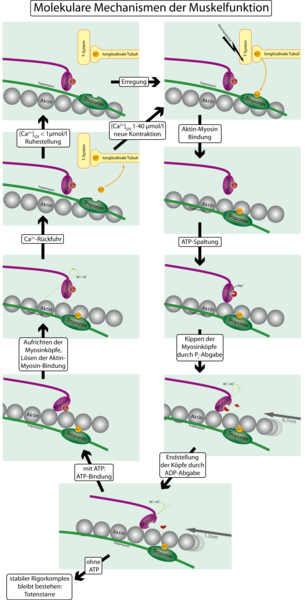
Molecular mechanisms of muscular function
