Seminal vesicle
Template:Infobox Anatomy
The seminal vesicles are a pair of simple tubular glands posteroinferior to the urinary bladder of males.
Anatomy
They are approximately 5 cm in length, though the full length of the gland is approximately 10 cm and curled up inside of the structure. Both glands form as an outpocketing of the wall of the ampulla of the vas deferens.
The ducts of the seminal vesicles open into the vas deferens as it enters the prostate gland.
Function
They secrete a significant proportion of the fluid that ultimately becomes semen. About 70% of the seminal fluid in humans originates from the seminal vesicles.
The thick secretions contain proteins, enzymes, fructose, mucus, vitamin C, flavins, phosphorylcholine and prostaglandins. The high fructose concentrations provide nutrient energy for the spermatozoa as they travel through the female reproductive system. The fluid is expelled under sympathetic contraction of the muscularis muscle coat.
Histology
Histologically, the seminal vesicles are defined by their tortuous pathways, false lumens, pseudo-stratified columnar epithelium and cuboidal cells along the basal layer.
The height of these columnar cells, and therefore activity, is dependent upon testosterone levels in the blood.
The lumen is large and stores the fluid secretions between ejaculations.
The mucosa is arranged into convoluted folds, significantly increasing surface area.
Joining the epithelium to the underlying muscularis is a fibroelastic connective tissue layer, with the well-developed muscularis layer composed of an inner circular and outer longitudinal layer of smooth muscle.
Additional images
-
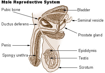
Male reproductive system.
-
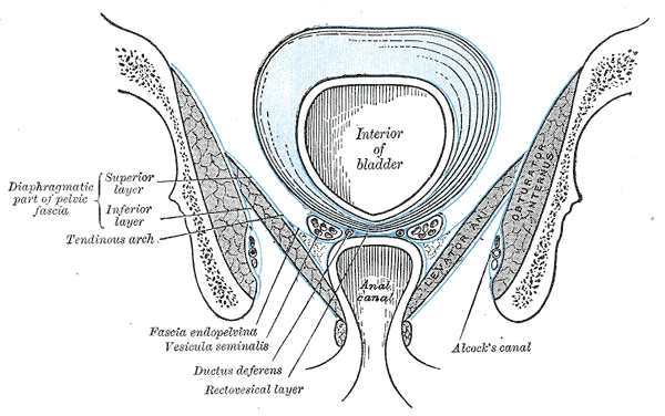
Coronal section of pelvis, showing arrangement of fasciæ. Viewed from behind.
-
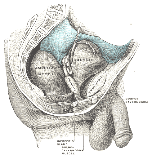
Male pelvic organs seen from right side.
-
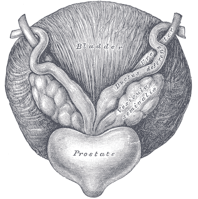
Fundus of the bladder with the vesiculæ seminales.
-
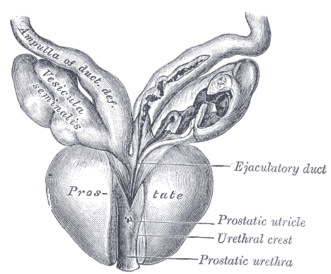
Vesiculæ seminales and ampullæ of ductus deferentes, seen from the front.
-
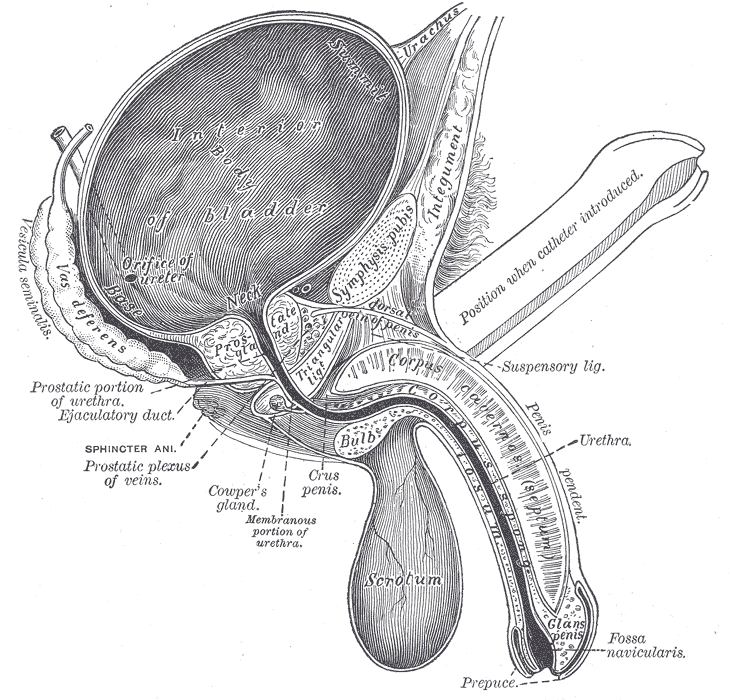
Vertical section of bladder, penis, and urethra.
-
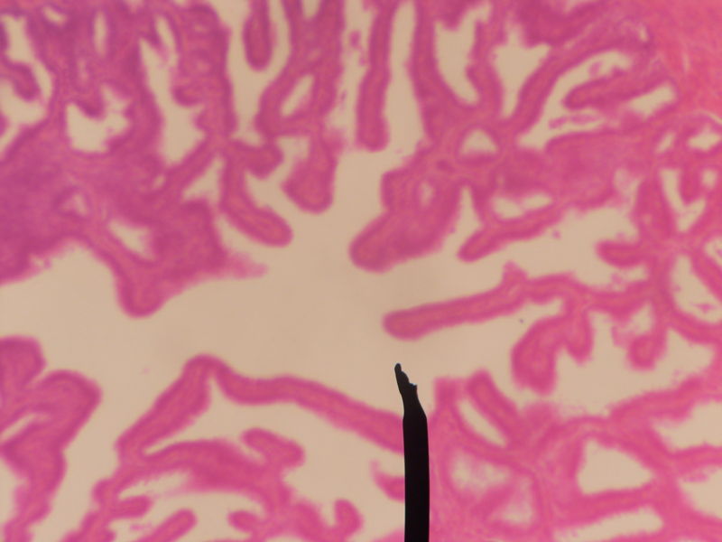
Cross section of seminal vesicle through a microscope.


External links
- Histology image: 17501loa – Histology Learning System at Boston University - "Male Reproductive System: prostate, seminal vesicle"
- Template:SUNYAnatomyLabs - "The Male Pelvis: The Urinary Bladder"
- Template:SUNYAnatomyLabs - "The Male Pelvis: Structures Located Posterior to the Urinary Bladder"
Template:Male reproductive system
cs:Semenný váček da:Sædblære de:Samenblase id:Vesikula seminalis it:Vescicola seminale lt:Sėklinės pūslelės nl:Zaadblaas simple:Seminal vesicle sk:Semenný mechúrik fi:Rakkularauhanen sv:Sädesblåsa