Pyonephrosis pathophysiology
|
Pyonephrosis Microchapters |
|
Diagnosis |
|---|
|
Treatment |
|
Case Studies |
|
Pyonephrosis pathophysiology On the Web |
|
American Roentgen Ray Society Images of Pyonephrosis pathophysiology |
|
Risk calculators and risk factors for Pyonephrosis pathophysiology |
Editor-In-Chief: C. Michael Gibson, M.S., M.D. [1]; Associate Editor(s)-in-Chief: Harsh Vardhan Chawla, M.B.B.S.[2]
Overview

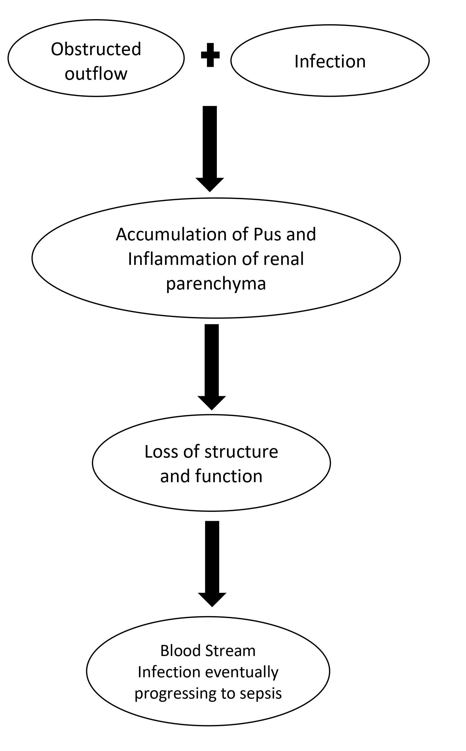
Pyonephrosis can be seen as a complication of acute pyelonephritis, usually seen with complete or incomplete obstruction of tubules. Obstruction of ureter and renal pelvis causes dilatation of tubular system which in turn leads to hydronephrosis. The dilatation of the tubular system serves as a nidus for infection because the pathogens multiply easily in obstructed and dilated tubules leading to suppurative inflammation.
Pathophysiology
- Pyonephrosis can be seen as a complication of acute pyelonephritis, usually seen with complete or incomplete obstruction of tubules[1].
- Obstruction of ureter and renal pelvis causes dilatation of tubular system which in turn leads to hydronephrosis.
- The dilatation of the tubular system serves as a nidus for infection because the pathogens multiply easily in obstructed and dilated tubules leading to suppurative inflammation.
- The exudates fill the tubules, renal pelvis, calyces, and ureter with pus that is difficult to drain.
- The accumulation of pus in the tubules eventually leads to structural and functional loss of the renal parenchyma which can cause a complete or incomplete loss of function initially.[2]
- The combination of obstruction with the infections may rapidly progress to sepsis.
- Therefore, it becomes critical to promptly diagnose and treat the condition to prevent renal loss of function and bloodstream infection leading to sepsis.
References
- ↑ Kumar, Vinay (2015). Robbins and Cotran pathologic basis of disease. Philadelphia, PA: Elsevier/Saunders. ISBN 978-1-4557-2613-4.
- ↑ Tamburrini S, Lugarà M, Iannuzzi M, Cesaro E, De Simone F, Del Biondo D; et al. (2021). "Pyonephrosis Ultrasound and Computed Tomography Features: A Pictorial Review". Diagnostics (Basel). 11 (2). doi:10.3390/diagnostics11020331. PMC 7921924 Check
|pmc=value (help). PMID 33671431 Check|pmid=value (help).