Hemorrhagic stroke MRI
|
Hemorrhagic stroke Microchapters |
|
Diagnosis |
|---|
|
Treatment |
|
AHA/ASA Guidelines for the Management of Spontaneous Intracerebral Hemorrhage (2015) |
|
AHA/ASA Guidelines for the Management of Aneurysmal Subarachnoid Hemorrhage (2012) |
|
AHA/ASA Guideline Recommendation for the Primary Prevention of Stroke (2014) |
|
AHA/ASA Guideline Recommendations for Prevention of Stroke in Women (2014) Sex-Specific Risk Factors
Risk Factors Commoner in Women |
|
Case Studies |
|
Hemorrhagic stroke MRI On the Web |
|
American Roentgen Ray Society Images of Hemorrhagic stroke MRI |
|
Risk calculators and risk factors for Hemorrhagic stroke MRI |
Editor-In-Chief: C. Michael Gibson, M.S., M.D. [1]; Associate Editor(s)-in-Chief: Sara Mehrsefat, M.D. [2]
Overview
MRI is better than CT for detection of acute ischaemia, and also detection of acute and chronic hemorrhage. Therefore it should be the preferred test for accurate diagnosis of patients with suspected acute stroke.[1]
MRI
MRI is better than CT for detection of acute ischaemia, and also detection of acute and chronic hemorrhage. Therefore it should be the preferred test for accurate diagnosis of patients with suspected acute stroke.[1]
- T2 susceptibility-weighted MRI are as sensitive as CT for detection of acute blood and are more sensitive for identification of prior hemorrhage.[1]
Images
The following are images associated with hemorrhagic stroke involves the left lentiform nucleus and internal capsule.[2]
-
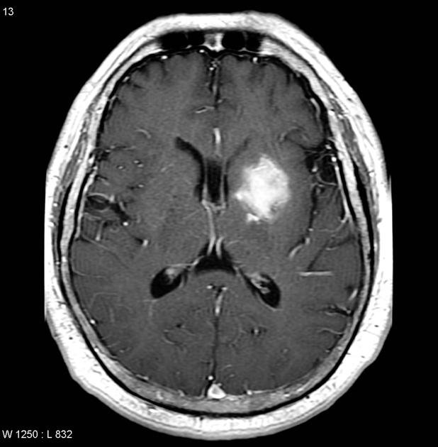
Axial T1
-
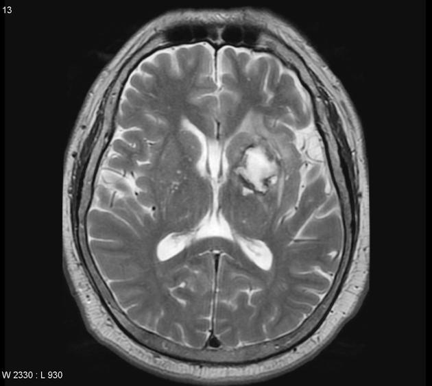
T2
-

Sagittal T1 C+


References
- ↑ 1.0 1.1 1.2 Chalela JA, Kidwell CS, Nentwich LM, Luby M, Butman JA, Demchuk AM; et al. (2007). "Magnetic resonance imaging and computed tomography in emergency assessment of patients with suspected acute stroke: a prospective comparison". Lancet. 369 (9558): 293–8. doi:10.1016/S0140-6736(07)60151-2. PMC 1859855. PMID 17258669.
- ↑ Intracerebral Hemotrrhage https://radiopaedia.org/cases/intracerebral-haemorrhage-2 Accessed on November 9, 2016