First degree AV block electrocardiogram
|
First degree AV block Microchapters |
|
Diagnosis |
|---|
|
Treatment |
|
Case Studies |
|
First degree AV block electrocardiogram On the Web |
|
American Roentgen Ray Society Images of First degree AV block electrocardiogram |
|
Risk calculators and risk factors for First degree AV block electrocardiogram |
Editor-In-Chief: C. Michael Gibson, M.S., M.D. [1]; Associate Editor(s)-in-Chief: Cafer Zorkun, M.D., Ph.D. [2]
Overview
In normal individuals, the AV node slows the conduction of electrical impulse through the heart. This is manifest on a surface EKG as the PR interval. The normal PR interval is from 120 milliseconds (ms) to 200 milliseconds (ms) in duration. This is measured from the initial deflection of the P wave to the beginning of the QRS complex.
In first degree heart block, the diseased AV node conducts the electrical activity slower. This is seen as a PR interval greater than 200 milliseconds (ms) in length on the surface EKG. It is usually an incidental finding on a routine EKG.
First degree heart block does not require any particular evaluation except for electrolyte and drug screens especially if an overdose is suspected.
Electrocardiogram
- ECG findings in patients with first degree heart block include the following:[1]
- PR interval is greater than 0.20 seconds = 200 miliseconds
- Each P wave is followed by a QRS
- Range of PR interval is between 0.21 and 0.40 seconds
- P wave may be mistaken for a T wave or a U wave
- The PR interval is more variable in those without heart disease
- In patients with a narrow QRS, His bundle recordings show that the conduction delay is in the AV node, with prolongation of the atrial His (AH) time, rarely is a prolonged His ventricular (HV) time responsible.
- In patients with PR prolongation and QRS prolongation, then the conduction delay may occur in various regions of the conduction system
EKG Examples
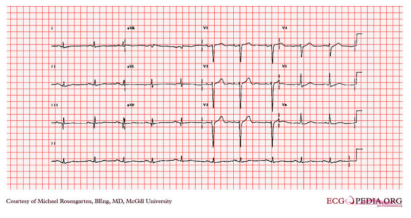
Shown below is an image of an electrocardiogram of a 64 year-old man with a history of coronary artery disease. The patient was taking amiodarone, metoprolol, and vasotec at the time of this recording. This was a routine recording. The electrocardiogram shows sinus rhythm with a prolonged P wave > 120 milliseconds. The P wave is notched in the inferior leads. This is consistent with a left atrial abnormality. The PR interval is long at 200 milliseconds and is diagnostic of first-degree heart block. The Q waves in the inferior leads suggest an inferior wall infarction.

Copyleft image obtained courtesy of ECGpedia, http://en.ecgpedia.org/wiki/Main_Page
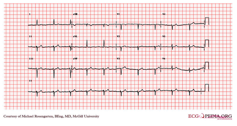
Shown below is the electrocardiogram of a 77 year old man with a history of coronary artery disease. He was taking Monopril, metoprolol, ASAand Hytrin. The electrocardiogram shows sinus rhythm with a marked first degree heart block ( about 360ms). There is also a poor R wave progression across the precordial leads and a Q wave in V2 suggestive of a previousanterior wall infarction. The QRS also has a left axis deviation best described as a left anterior hemi-block.

Copyleft image obtained courtesy of ECGpedia, http://en.ecgpedia.org/wiki/Main_Page
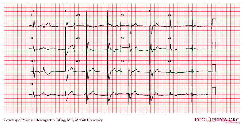
Shown below is an electrocardiogram from a 87 year old man with a history of atrial fibrillation. His medications were coumadin and Monopril. The cardiogram shows sinus rhythm with rate of about 50/min, and a marked first degree heart block with a PR interval of about 350 msec.
The first complex on the left is a fusion between the patient's native QRS and the pacemaker spike (this is normal operation) this is followed by a PVC. Note the small blip following the PVC is artifact and is not a failure to capture of the pacemaker. The pacemaker is working well as a VVI pacer set at 50/min. The large spikes suggest a unipolar lead.

Copyleft image obtained courtesy of ECGpedia, http://en.ecgpedia.org/wiki/Main_Page
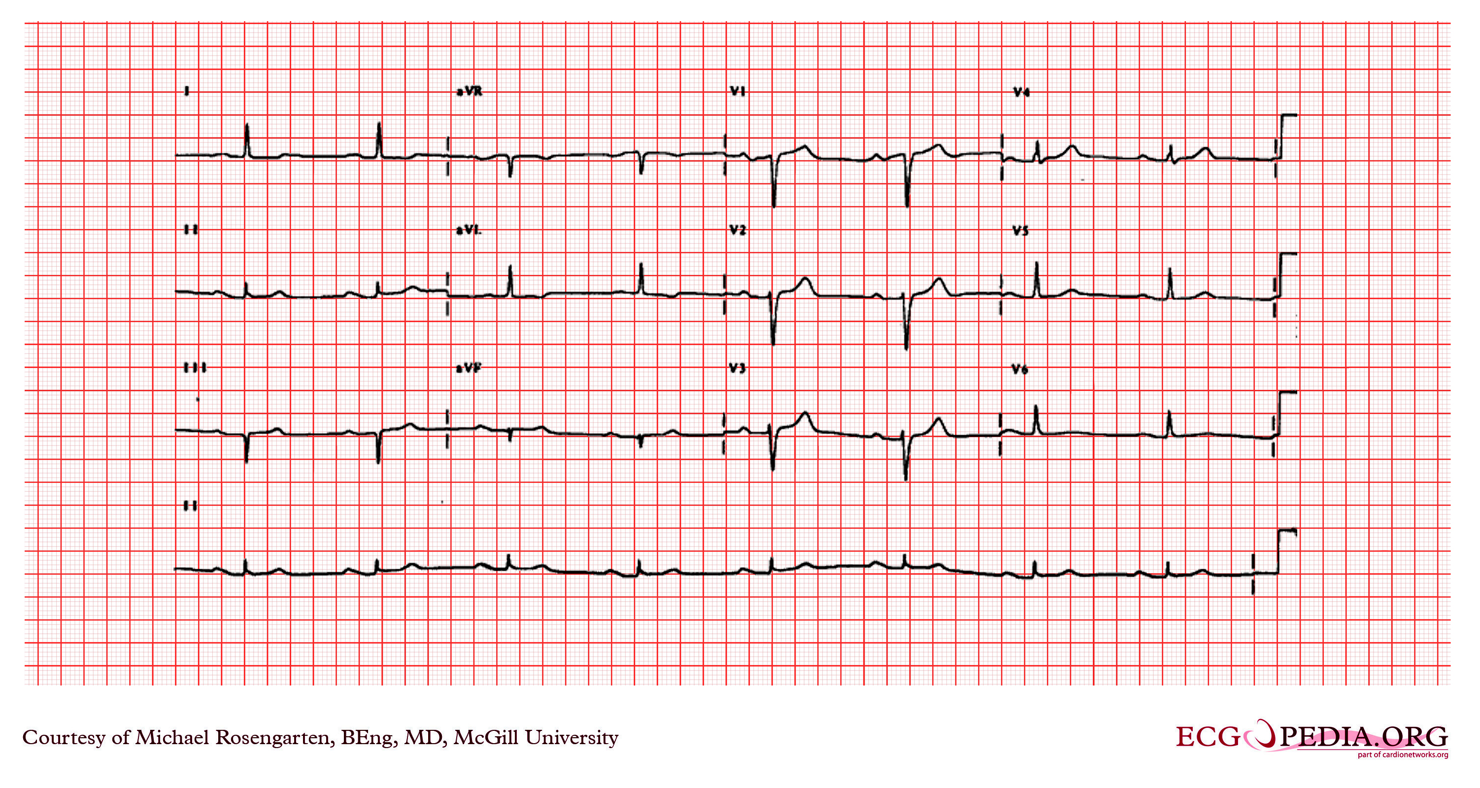
Shown below is an electrocardiogram demonstrating sinus rhythm with first degree heart block (PR segment > 200ms.) Note the P waves in the inferior leads are greater than 120 ms. in duration, which suggests left atrial abnormality.

Copyleft image obtained courtesy of ECGpedia, http://en.ecgpedia.org/wiki/Main_Page
For more EKG examples of First Degree AV Block click here
References
- ↑ Barold SS (1996). "Indications for permanent cardiac pacing in first-degree AV block: class I, II, or III?". Pacing Clin Electrophysiol. 19 (5): 747–51. doi:10.1111/j.1540-8159.1996.tb03355.x. PMID 8734740.