File:Ultrasound imaging of ovarian thecofibroma.jpg
Summary
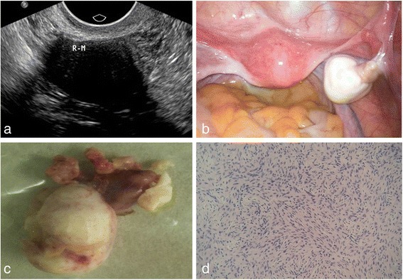
A 53-year-old woman with a pelvic mass discovered by routing physical examination. a On ultrasound examination, a 28 mm*22 mm*26 mm well-circumscribed hypoechoic mass was observed in the right ovary, with posterior echo attenuation. b On laparoscopy surgery, right ovarian was hard and enlarged, the surface smooth, having a good mobility; in the pelvic, a small number of pale yellow ascites were observed. c Pathologically, right ovarian was enlarged, with tough textures and grayish white surfaces. d Pathological findings confirmed thecofibroma in right ovarian,Chen H, Liu Y, Shen LF, Jiang MJ, Yang ZF, Fang GP. Ovarian thecoma-fibroma groups: clinical and sonographic features with pathological comparison. J Ovarian Res. 2016;9(1):81. Published 2016 Nov 22. doi:10.1186/s13048-016-0291-2,https://www.ncbi.nlm.nih.gov/pmc/articles/PMC5120502/
File history
Click on a date/time to view the file as it appeared at that time.
| Date/Time | Thumbnail | Dimensions | User | Comment | |
|---|---|---|---|---|---|
| current | 19:21, 19 April 2019 | | 567 × 394 (88 KB) | Maneesha Nandimandalam (talk | contribs) | A 53-year-old woman with a pelvic mass discovered by routing physical examination. a On ultrasound examination, a 28 mm*22 mm*26 mm well-circumscribed hypoechoic mass was observed in the right ovary, with posterior echo attenuation. b On laparoscopy su... |
You cannot overwrite this file.
File usage
The following page uses this file: