File:Squamous cell Ca.jpg
Jump to navigation
Jump to search
Size of this preview: 523 × 600 pixels. Other resolution: 600 × 688 pixels.
Original file (600 × 688 pixels, file size: 197 KB, MIME type: image/jpeg)
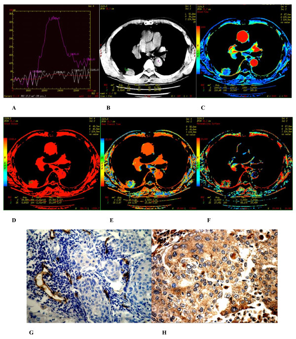
(A-H) Hamartoma found in apicoposterior segment of superior of the left lung of a 54-year-old male. (A) Time density curve. (B-F) (original image, BF, BV, MTT, PS) type I parametric maps, PS value was moderate (12.029). (G) CD34 staining shows a few of more mature microvessels (× 200). (H) VEGF expression is negative (× 400).
File history
Click on a date/time to view the file as it appeared at that time.
| Date/Time | Thumbnail | Dimensions | User | Comment | |
|---|---|---|---|---|---|
| current | 00:06, 17 February 2018 | | 600 × 688 (197 KB) | Dildar Hussain (talk | contribs) | (A-H) Hamartoma found in apicoposterior segment of superior of the left lung of a 54-year-old male. (A) Time density curve. (B-F) (original image, BF, BV, MTT, PS) type I parametric maps, PS value was moderate (12.029). (G) CD34 staining shows a few of... |
You cannot overwrite this file.
File usage
The following 4 pages use this file: