File:Phlebovirus06.jpeg
Jump to navigation
Jump to search
Size of this preview: 395 × 600 pixels. Other resolution: 700 × 1,063 pixels.
Original file (700 × 1,063 pixels, file size: 139 KB, MIME type: image/jpeg)
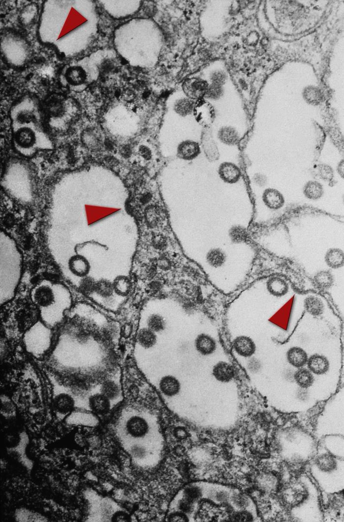
Under a very high magnification, this transmission electron micrograph (TEM) revealed some of the ultrastructural morphology seen in an unknown tissue sample, which had been caused by the spherical-shaped, enveloped Rift Valley fever (RVF) virus. In this particular view you can see some virions budding from the cell membrane, indicated by arrowheads.
File history
Click on a date/time to view the file as it appeared at that time.
| Date/Time | Thumbnail | Dimensions | User | Comment | |
|---|---|---|---|---|---|
| current | 16:34, 11 December 2014 | | 700 × 1,063 (139 KB) | Jesus Hernandez (talk | contribs) | Under a very high magnification, this transmission electron micrograph (TEM) revealed some of the ultrastructural morphology seen in an unknown tissue sample, which had been caused by the spherical-shaped, enveloped Rift Valley fever (RVF) virus. In th... |
You cannot overwrite this file.
File usage
The following file is a duplicate of this file (more details):
The following page uses this file: