File:PET-CT scan.jpg
PET-CT_scan.jpg (472 × 518 pixels, file size: 79 KB, MIME type: image/jpeg)
Summary
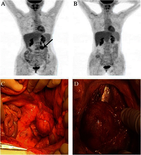
PET-CT scan and macroscopic findings in a recurrent patient (case 11). (A) PET-CT scan before treatment; a black arrow points to the metastatic tumor in the left portion of the fourth lumbar vertebra. (B) PET-CT scan after treatment. (C) and (D) show the retroperitoneal tumor fused by several para-aortic lymph node,Qian Q, You Y, Yang J, et al. Management and prognosis of patients with ovarian sex cord tumor with annular tubules: a retrospective study. BMC Cancer. 2015;15:270. Published 2015 Apr 12. doi:10.1186/s12885-015-1277-y,https://www.ncbi.nlm.nih.gov/pmc/articles/PMC4408581/
File history
Click on a date/time to view the file as it appeared at that time.
| Date/Time | Thumbnail | Dimensions | User | Comment | |
|---|---|---|---|---|---|
| current | 17:25, 17 April 2019 | | 472 × 518 (79 KB) | Maneesha Nandimandalam (talk | contribs) | PET-CT scan and macroscopic findings in a recurrent patient (case 11). (A) PET-CT scan before treatment; a black arrow points to the metastatic tumor in the left portion of the fourth lumbar vertebra. (B) PET-CT scan after treatment. (C) and (D) show t... |
You cannot overwrite this file.
File usage
The following page uses this file: