File:Neurovascular compression of the trigeminal root.jpeg

Original file (506 × 1,280 pixels, file size: 112 KB, MIME type: image/jpeg)
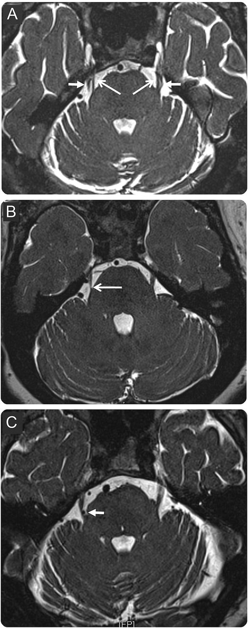
3D constructive interference in steady state MRI shows axial sections at the level of trigeminal nerve root entry into the pons. (A) Bilateral neurovascular contact without morphologic changes of the root in a patient with left trigeminal neuralgia (TN). Nerve (long arrows) and blood vessel (short arrows) appear hypointense surrounded by hyperintense CSF. Contact is seen at the root entry zone as well as mid-cisternal segment. (B, C) Morphologic changes exceeding mere neurovascular contact of the trigeminal nerve root are compatible with the diagnosis of classical TN. (B) Root atrophy in a patient with right TN. (C) Indentation and dislocation of the root in a patient with right TN (short arrow).
File history
Click on a date/time to view the file as it appeared at that time.
| Date/Time | Thumbnail | Dimensions | User | Comment | |
|---|---|---|---|---|---|
| current | 19:38, 23 June 2018 | 506 × 1,280 (112 KB) | Simraa.K (talk | contribs) | 3D constructive interference in steady state MRI shows axial sections at the level of trigeminal nerve root entry into the pons. (A) Bilateral neurovascular contact without morphologic changes of the root in a patient with left trigeminal neuralgia (TN... |
You cannot overwrite this file.
File usage
The following 2 pages use this file: