File:MRI pilocytic astrocytoma 1.jpg
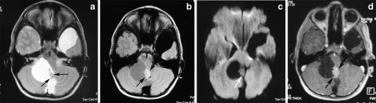
A large, cystic cerebellar pilocytic astrocytoma in a 7-year-old boy. a An axial T2-weighted image shows a hyperintense mass of the right cerebellar hemisphere with a less intense soft-tissue nodule along its medial margin and without surrounding oedema (arrows). Note the arachnoid cyst in the left middle cranial fossa. b On an axial FLAIR image, the cystic component shows low signal intensity that is higher than that of the CSF, and the soft tissue nodule is homogeneous and slightly hyperintense (arrows). c On a diffusion-weighted image, the mural nodule appears isointense (arrow). d An axial, contrast-enhanced, T1-weighted image demonstrates intense enhancement of the mural nodule (arrows).
File history
Click on a date/time to view the file as it appeared at that time.
| Date/Time | Thumbnail | Dimensions | User | Comment | |
|---|---|---|---|---|---|
| current | 17:34, 29 October 2015 | 547 × 150 (27 KB) | Sujit Routray (talk | contribs) | A large, cystic cerebellar pilocytic astrocytoma in a 7-year-old boy. '''a''' An axial T2-weighted image shows a hyperintense mass of the right cerebellar hemisphere with a less intense soft-tissue nodule along its medial margin and without surrounding... | |
| 17:34, 29 October 2015 | 547 × 150 (27 KB) | Sujit Routray (talk | contribs) | A large, cystic cerebellar pilocytic astrocytoma in a 7-year-old boy. '''a''' An axial T2-weighted image shows a hyperintense mass of the right cerebellar hemisphere with a less intense soft-tissue nodule along its medial margin and without surrounding... |
You cannot overwrite this file.
File usage
There are no pages that use this file.