File:IJRI-25-109-g016.jpg
Jump to navigation
Jump to search
No higher resolution available.
IJRI-25-109-g016.jpg (539 × 219 pixels, file size: 26 KB, MIME type: image/jpeg)
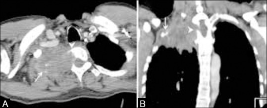
Superior sulcus tumor. Axial (A) and coronal (B) CT scans show a large mass in the apex of the right lung causing destruction of the first and second ribs (arrows) with erosion of the right half of the vertebral body (arrowheads) suggestive of a superior sulcus tumor
File history
Click on a date/time to view the file as it appeared at that time.
| Date/Time | Thumbnail | Dimensions | User | Comment | |
|---|---|---|---|---|---|
| current | 18:13, 16 February 2018 | 539 × 219 (26 KB) | Dildar Hussain (talk | contribs) | Superior sulcus tumor. Axial (A) and coronal (B) CT scans show a large mass in the apex of the right lung causing destruction of the first and second ribs (arrows) with erosion of the right half of the vertebral body (arrowheads) suggestive of a superi... |
You cannot overwrite this file.
File usage
The following 4 pages use this file: