File:IJRI-25-109-g008.jpg
Jump to navigation
Jump to search
No higher resolution available.
IJRI-25-109-g008.jpg (538 × 423 pixels, file size: 47 KB, MIME type: image/jpeg)
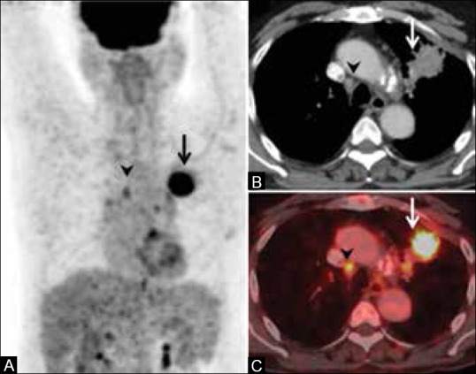
FDG PET in nodal disease. Maximum intensity projection (MIP) image shows an FDG-avid primary lung tumor on the left side (arrow, A) and a focus of FDG uptake in the mediastinum (arrowhead, A). CT scan shows enhancing, spiculated primary tumor (arrow, B) and a small right paratracheal node (arrowhead, B) which is negative by size criteria. Fused PET/CT image shows FDG concentration in the primary (arrow, C) as well as the node (arrowhead, C), suggesting metastatic involvement. Mediastinoscopy and biospy revealed metastatic node-N3 disease
File history
Click on a date/time to view the file as it appeared at that time.
| Date/Time | Thumbnail | Dimensions | User | Comment | |
|---|---|---|---|---|---|
| current | 16:18, 16 February 2018 | | 538 × 423 (47 KB) | Dildar Hussain (talk | contribs) | FDG PET in nodal disease. Maximum intensity projection (MIP) image shows an FDG-avid primary lung tumor on the left side (arrow, A) and a focus of FDG uptake in the mediastinum (arrowhead, A). CT scan shows enhancing, spiculated primary tumor (arrow, B... |
You cannot overwrite this file.
File usage
The following page uses this file: