File:HIV opportunistic10.jpeg
HIV_opportunistic10.jpeg (700 × 467 pixels, file size: 20 KB, MIME type: image/jpeg)
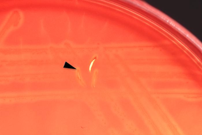
Magnified 100x, this 1977 photograph depicted a Petri dish filled with trypticase soy agar medium containing 5% defibrinated sheep's blood, i.e., blood agar plate (BAP). After having been inoculated with alpha-hemolytic Streptococcus anginosus bacteria, i.e., a member of the Gram-positive viridans group of streptococci (VGS), using a streak/stab technique, the BAP was incubated in a carbon dioxide enriched atmosphere at 35oC for 24 hours. The culture grew bacterial colonies. Typical alpha-hemolytic colonies were seen adjacent to the stab sites (arrow). The hazy, faded, indistinct region surrounding each colony indicated that red blood cells (RBCs) were destroyed, or "hemolyzed", in the BAP, and that these bacteria were indeed alpha-hemolytic in nature.
It is the incomplete nature of the hemolytic reaction adjacent to the colonies, which spares numbers of RBCs in the blood agar medium, that is of qualitative importance when distinguishing alpha from beta-hemolysis.
File history
Click on a date/time to view the file as it appeared at that time.
| Date/Time | Thumbnail | Dimensions | User | Comment | |
|---|---|---|---|---|---|
| current | 21:20, 19 November 2014 | | 700 × 467 (20 KB) | Jesus Hernandez (talk | contribs) | Magnified 100x, this 1977 photograph depicted a Petri dish filled with trypticase soy agar medium containing 5% defibrinated sheep's blood, i.e., blood agar plate (BAP). After having been inoculated with alpha-hemolytic Streptococcus anginosus bacteria... |
You cannot overwrite this file.
File usage
The following page uses this file: