File:HIV lab01.jpeg
Jump to navigation
Jump to search
No higher resolution available.
HIV_lab01.jpeg (700 × 525 pixels, file size: 48 KB, MIME type: image/jpeg)
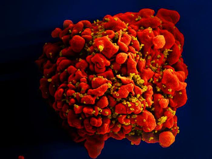
Produced by the National Institute of Allergy and Infectious Diseases (NIAID), this digitally-colorized scanning electron micrograph (SEM) depicts a single, red-colored H9-T cell that had been infected by numerous, spheroid-shaped, mustard-colored human immunodeficiency virus (HIV) particles, which can be seen sttached to the cell's surface membrane.
File history
Click on a date/time to view the file as it appeared at that time.
| Date/Time | Thumbnail | Dimensions | User | Comment | |
|---|---|---|---|---|---|
| current | 20:44, 19 November 2014 | | 700 × 525 (48 KB) | Jesus Hernandez (talk | contribs) | Produced by the National Institute of Allergy and Infectious Diseases (NIAID), this digitally-colorized scanning electron micrograph (SEM) depicts a single, red-colored H9-T cell that had been infected by numerous, spheroid-shaped, mustard-colored huma... |
You cannot overwrite this file.
File usage
The following page uses this file: