File:Entamoeba histolytica02.jpeg
Jump to navigation
Jump to search
Size of this preview: 636 × 600 pixels. Other resolution: 700 × 660 pixels.
Original file (700 × 660 pixels, file size: 51 KB, MIME type: image/jpeg)
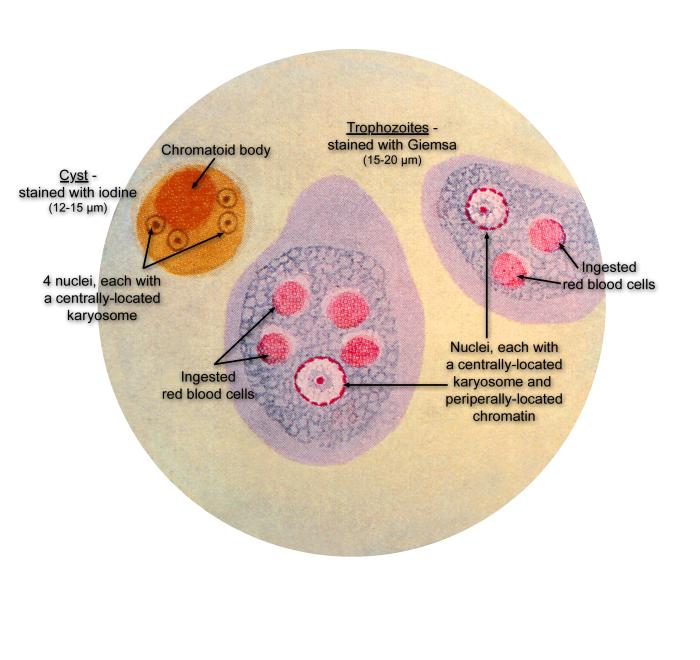
This illustration of a composite photomicrograph reveals the ultrastructural details seen in two stages of the life cycle of the parasite Entamoeba histolytica, the cystic stage (lt), stained with iodine, and the Giemsa-stained vegetative, trophozoite stage.
File history
Click on a date/time to view the file as it appeared at that time.
| Date/Time | Thumbnail | Dimensions | User | Comment | |
|---|---|---|---|---|---|
| current | 19:44, 14 November 2014 | | 700 × 660 (51 KB) | Jesus Hernandez (talk | contribs) | This illustration of a composite photomicrograph reveals the ultrastructural details seen in two stages of the life cycle of the parasite Entamoeba histolytica, the cystic stage (lt), stained with iodine, and the Giemsa-stained vegetative, trophozoite ... |
You cannot overwrite this file.
File usage
The following page uses this file: