File:Borrelia29.jpeg
Borrelia29.jpeg (700 × 475 pixels, file size: 43 KB, MIME type: image/jpeg)
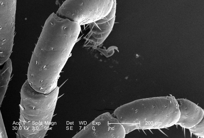
Under a relatively low magnification of 95X, this scanning electron micrograph (SEM) provided a closer view of this male Dermacentor sp. tick found upon a cat in the suburbs of Decatur, Georgia, which measured approximately 3.5mm from its gnathosoma (i.e., capitulum), which is where its mouthparts are located, to the distal abdominal margin (PHIL 9961). PHIL 9959 revealed all this tick’s legs, placing it into the Phylum Arthropoda, i.e., from jointed ( “Arthro”), and legs (“poda”), as well as the Class Arachnida, for they’ve eight of these legs, unlike insects, which use six appendages to move about. From proximal to distal location, each leg is comprised of a coxa, trochanter 1, trochanter 2, a femur, patella, tibia, a two-sectioned tarsus, and a two-part pretarsus, i.e., a pulvillus and claw. Here we see the femur, patella, and tibia of arachnid’s left 2nd and 3rd legs.
File history
Click on a date/time to view the file as it appeared at that time.
| Date/Time | Thumbnail | Dimensions | User | Comment | |
|---|---|---|---|---|---|
| current | 15:16, 26 November 2014 | | 700 × 475 (43 KB) | Jesus Hernandez (talk | contribs) | Under a relatively low magnification of 95X, this scanning electron micrograph (SEM) provided a closer view of this male Dermacentor sp. tick found upon a cat in the suburbs of Decatur, Georgia, which measured approximately 3.5mm from its gnathosoma (i... |
You cannot overwrite this file.
File usage
The following file is a duplicate of this file (more details):
The following page uses this file: