File:628px-Journal.pone.0008247.g001.png
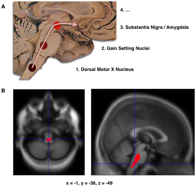
A. Schematic initial progression of Lewy body deposits in the first stages of Parkinson's Disease, as proposed by Braak and colleagues. B. Localization of the cluster of significant volume reduction in PD compared with HC. The significant cluster located in the medulla oblongata/pons is superimposed as a red blob on the mean normalized anatomical scan of all participants. The axial and sagital sections are centered on the peak of significance (−1; −36; −49).
Image comes from an article by Jubault et al. in PlosOne (2009) using Voxel Based Morphometry which searched for regional atrophy in idiopathic Parkinson's Disease by comparing a group of subjects with the disease and a group of healthy controls.
Citation of the original article: Jubault T, Brambati SM, Degroot C, Kullmann B, Strafella AP, et al. (2009) Regional Brain Stem Atrophy in Idiopathic Parkinson's Disease Detected by Anatomical MRI. PLoS ONE 4(12): e8247. doi:10.1371/journal.pone.0008247 DOI of the image: 10.1371/journal.pone.0008247.g001
File history
Click on a date/time to view the file as it appeared at that time.
| Date/Time | Thumbnail | Dimensions | User | Comment | |
|---|---|---|---|---|---|
| current | 16:22, 7 April 2018 | | 628 × 599 (255 KB) | Fahimeh Shojaei (talk | contribs) | A. Schematic initial progression of Lewy body deposits in the first stages of Parkinson's Disease, as proposed by Braak and colleagues. B. Localization of the cluster of significant volume reduction in PD compared with HC. The significant cluster locat... |
You cannot overwrite this file.
File usage
The following page uses this file: