D-dimer physiology
|
D-Dimer Microchapters |
|
Clinical Correlation |
|---|
|
Clinical Trials |
|
D-dimer physiology On the Web |
|
American Roentgen Ray Society Images of D-dimer physiology |
Editor-In-Chief: C. Michael Gibson, M.S., M.D. [1]
Overview
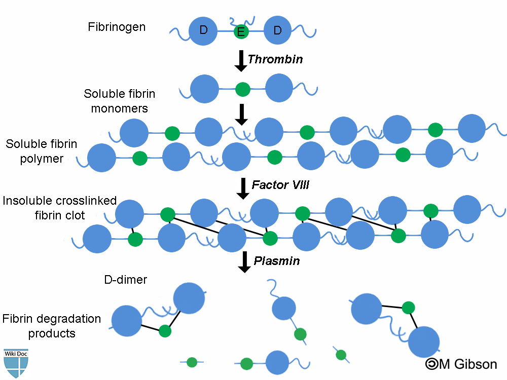
D-dimer is fibrin degradation product (FDP) resulting from the sequential break down of fibrin by the enzymes thrombin, factor VIII and plasmin.
Physiology
Fibrin degradation products (FDPs) are formed whenever fibrin is broken down by enzymes. In fact, FDP are formed as a result of the sequential actions of the following three different enzymes: thrombin, factor VIII and plasmin. Determining FDPs is not considered useful, as this does not indicate whether the fibrin is part of a blood clot (or being generated as part of inflammation).
D-dimers are unique in that they are the breakdown products of a fibrin mesh that has been stabilized by Factor XIII. This factor crosslinks the E-element to two D-elements. This is the final step in the generation of a thrombus.
Plasmin is a fibrinolytic enzyme that organizes clots and breaks down the fibrin mesh. It cannot, however, break down the bonds between one E and two D units. The protein fragment thus left over is a D-dimer.[1]
Shown below is an image summarizing the formation of D-dimers and other fibrin degradation products as a result of the sequential action of the three enzymes: thrombin, factor VIII and plasmin.

Genetics
Three genetic variants, F3, F5, and FGA, were associated with D-dimer levels accoding to data based on 13 cohorts involving 21 052 healthy individuals from 13 European ancestry. The locations of the three genetic variants F3, F5, and FGA are 1p22 1q24 and 4q32 respectively.[2]
References
- ↑ Adam SS, Key NS, Greenberg CS (2009). "D-dimer antigen: current concepts and future prospects". Blood. 113 (13): 2878–87. doi:10.1182/blood-2008-06-165845. PMID 19008457 Check
|pmid=value (help). - ↑ Smith NL, Huffman JE, Strachan DP, Huang J, Dehghan A, Trompet S; et al. (2011). "Genetic predictors of fibrin D-dimer levels in healthy adults". Circulation. 123 (17): 1864–72. doi:10.1161/CIRCULATIONAHA.110.009480. PMC 3095913. PMID 21502573.