Common hepatic duct
Editor-In-Chief: C. Michael Gibson, M.S., M.D. [1]
Overview
The common hepatic duct is the duct formed by the convergence of the right hepatic duct (which drains bile from the right functional lobe of the liver) and the left hepatic duct (which drains bile from the left functional lobe of the liver). The common hepatic duct then joins the cystic duct coming from the gallbladder to form the common bile duct.
Clinical significance
The hepatic duct transports more volume in people who have had their gallbladder removed.
The common hepatic duct has an important relationship with the right hepatic artery and the cystic artery. All of these must be identified during a cholecystectomy to avoid cutting or clipping the wrong structure.
Dimensions
Approximate length: 8 cm Approximate width: 8 mm
Additional images
-
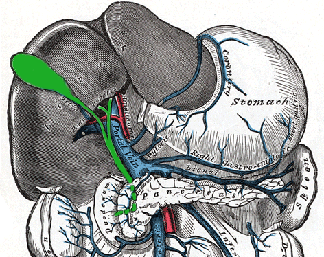
The portal vein and its tributaries.
-

External links
- Template:SUNYAnatomyLabs - "Stomach, Spleen and Liver: Contents of the Hepatoduodenal Ligament"
- Template:EMedicineDictionary
- Illustration