Atrioventricular septal defect classification
|
Atrioventricular septal defect Microchapters |
|
Differentiating Atrioventricular septal defect from other Diseases |
|---|
|
Diagnosis |
|
Treatment |
|
Atrioventricular septal defect classification On the Web |
|
American Roentgen Ray Society Images of Atrioventricular septal defect classification |
|
Risk calculators and risk factors for Atrioventricular septal defect classification |
Editor-In-Chief: C. Michael Gibson, M.S., M.D. [1]
Overview
Classification
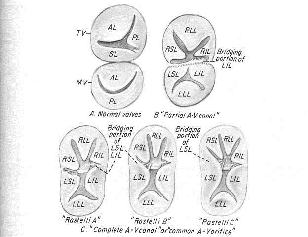
AVSD is classified as either complete, partial, or intermediate/transitional. Classification is based on the extent of loss of separation between the chambers of the heart. In partial AVSD, there is a defect in the primum or inferior part of the atrial septum but no direct intraventricular communication (ostium primum defect). In complete AVSD, there is a large ventricular component beneath either or both the superior or inferior bridging leaflets of the AV valve. In intermediate or transitional AVSD, the leaflets of the common AV valve are stuck to the ventricular septum causing a division of the two valves and resulting in a marginal sized hole between the ventricle. Transitional AVSDs are similar to partial AVSDs in behavior but visually appear more like a complete AVSD.
