Adductor magnus muscle
Overview
The adductor magnus is a large triangular muscle, situated on the medial side of the thigh.
The portion which arises from the ischiopubic ramus (a small part of the inferior ramus of the pubis, and the inferior ramus of the ischium) is called the "adductor portion", and the portion arising from the tuberosity of the ischium is called the "hamstring portion". The hamstring portion is not considered part of the hamstring group of muscles, but it is adjacent to it.
Adductor part
Those fibers which arise from the ramus of the pubis are short, horizontal in direction, and are inserted into the rough line leading from the greater trochanter to the linea aspera, medial to the gluteus maximus.
Those fibers from the ramus of the ischium are directed downward and lateralward with different degrees of obliquity, to be inserted, by means of a broad aponeurosis, into the linea aspera and the upper part of its medial prolongation below.
Hamstring part
The medial portion of the muscle, composed principally of the fibers arising from the tuberosity of the ischium, forms a thick fleshy mass consisting of coarse bundles which descend almost vertically, and end about the lower third of the thigh in a rounded tendon which is inserted into the adductor tubercle on the medial condyle of the femur, and is connected by a fibrous expansion to the line leading upward from the tubercle to the linea aspera.
Osseoaponeurotic openings
At the insertion of the muscle, there is a series of osseoaponeurotic openings, formed by tendinous arches attached to the bone. The upper four openings are small, and give passage to the perforating branches of the profunda femoris artery. The lowest is of large size, and transmits the femoral vessels to the popliteal fossa.
Action
The function of the adductor magnus is to serve as a powerful extensor of the thigh, along with being able to flex and laterally rotate the thigh.
See also
Additional images
-
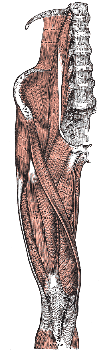 Muscles of the iliac and anterior femoral regions.
 -
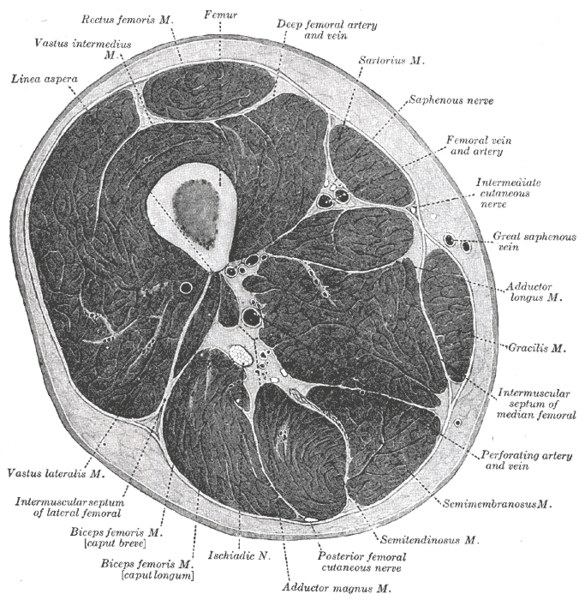 Cross-section through the middle of the thigh.
 -
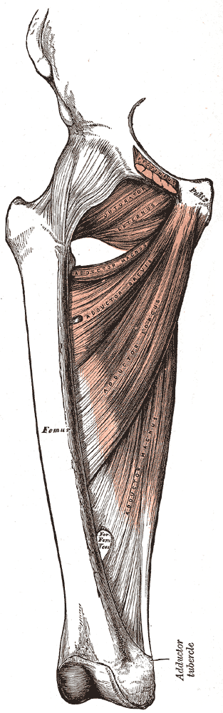 Deep muscles of the medial femoral region.
 -
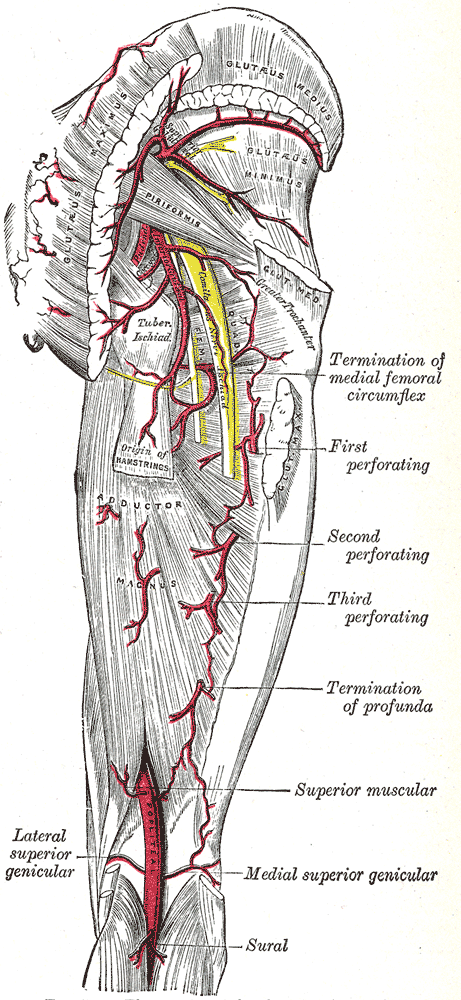 The arteries of the gluteal and posterior femoral regions.


References
External links
- Template:MuscleLoyola
- Template:GPnotebook
- Template:SUNYAnatomyLabs
- Template:EMedicineDictionary
- PTCentral
Template:Gray's Template:Muscles of lower limb